Scientific Classification
| Kingdom: Animalia |
| Phylum: Arthropoda |
| Class: Trilobita |
| Order: Phacopida |
| Family: Acastidae |
| Genus: Greenops |
| Species: Greenops barberi Lieberman & Kloc 1997 |
Information
Geological Range
Paleogeographic Distribution
Stratigraphic Occurrences
| Moscow Formation |
| Windom Member |
| Ludlowville Formation |
| Wanakah Member |
| Widder Formation |
References
Lieberman (1997): Evolutionary and biogeographic patterns in the Asteropyginae (Trilobita, Devinian) Delo, 1935. in Bulletin of the American Museum of Natural History.
Wilson (2014): p. 204.
Remarks
From Wilson (2014): “In the early studies of the asteropygine trilobites of New York (e.g., Hall & Clarke, 1888; Grabau, 1898), most specimens were classified and figured as one of two species. If the specimen had short, rounded pleural lappets, it was ‘Cryphaeus boothi Green,’ 1837 (later Greenops boothi). If it had long, spine-like kappets, it was “Cryphaeus calliteles” Green, 1837 (later Greenops calliteles)… size varies greatly, most specimens are ca. 25 mm. L, although specimens of up to 50mm or more are found…More recent studies (Lieberman & Kloc, 1997; Whiteley et al., 2002) have divided the New York “Greenops” into two genera, Greenops and Bellacartwrightia, each with several new species.”
See Bignon and Cronier (2014) for more information regarding the systematics of Greenops.
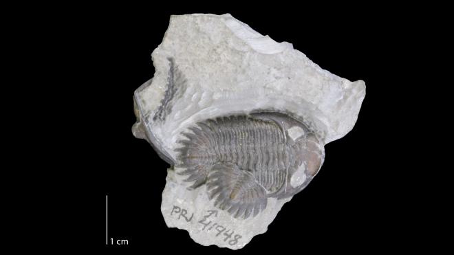
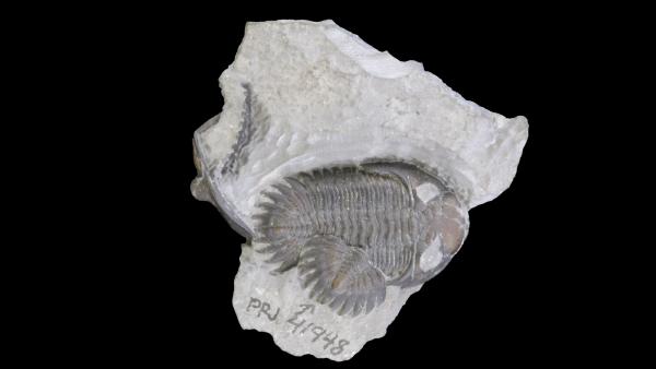
Description: From Lieberman (1997): “Cephalic length (sag.) 50% of width. Axial furrow diverges more strongly anterior to S1 than posterior to S1, diverges less strongly anterior to anterior half of L3, diverging forward at about 250, narrow, moderately incised. Cranidial anterior border developed as moderate lip deflected moderately anterior to facial suture. Preglabellar furrow shallow and narrow. Length (exsag., sag.) of cranidial anterior border increases slightly from lateral to medial edges of frontal lobe. Facial suture anterior to eyes flexes laterally then medially, with smoothly convex margin laterally. Posterior branch of facial suture flexes weakly anteriorly then posteriorly laterally. Glabellar length (sag.) equal to 100% of width (tr.) across frontal lobe; frontal lobe ellipsoid, about 55% length (sag.) of glabella; L1-L3 flat (sag.); frontal lobe at same elevation as that of posterior glabellar region; anterior part of frontal lobe moderately declined forward, smoothly rounded in dorsal view. PMI a rounded depression. S3 shallow, straight, anterolateral edge transverse, equally incised medially and distally, branches diverging at about 1400. Sagittal region of L3-L2 nearly flat (tr.), lateral lobes weakly declined abaxially…Genal spine developed as moderately long flange extending back to fifth thoracic segment, cephalic border furrow bisects spine; lateral and medial edges of genal spine evenly inclined; medial edge of genal spine not significantly thickened relative to lateral edge; lateral margin of genal spine parallels sagittal line, straight at posterior end; interior margin of genal spine evenly deflected laterally. Raised ridge on dorsal surface of genal spine at juncture of posterior border furrow and lateral border furrow forms smooth curve convex distally. Lateral border furrow narrow, shallow, lateral border weakly widens posteriorly. Anterior margin of cephalic doublure rounded. Prosopon of fine tubercles. Thorax of 11 segments. Axial ring about 25% of width (tr.) of thorax, of equal length (exsag., sag.), at distal ends and medially flexed forward. Anterior margin of ring well defined sagittally, inclined posterodorsally, most elevated near posterior edge; at lateral margin of axial rings, circular fenestrae present. Articulating half ring set slightly below axial ring…Pygidium broadly triangular in outline excluding marginal lappets, length about 60% of width without lappets, with five pairs of pleural lappets. Axial furrows shallow, narrow, converging at 250 angle anterior to fifth pygidial axial ring, roughly parallel posterior to fifth pygidial axial ring. Axis 30% of pygidial width (tr.) anteriorly, with 11 rings; rings of nearly equal length (exsag., sag.) distally and medially, longest medially; anterior edge of axial rings posterior to fifth axial ring from midline to distal edge convex anteriorly. Ring furrows moderately incised. Lateral margins of pygidial lappets curved, medial margins straight, distal tips pointed. Axial terminus triangular, does not project as far posteriorly as other lappets; postaxial region 15% of pygidial length (sag.). Pygidial pleural field flanking posterior part of pygidial axis faintly excavated; anterior and posterior bands of pleural segment equally elevated; tops of segments flat; pleural furrows deeper than interpleural furrows, short (exsag.). Laterally pygidial interpleural furrows arch at same angle as medially. In medial region of adjacent pleural segments, posterior region of anterior segment equal in length (exsag.) to anterior part of posterior segment.”
Online Resources
Media
Images